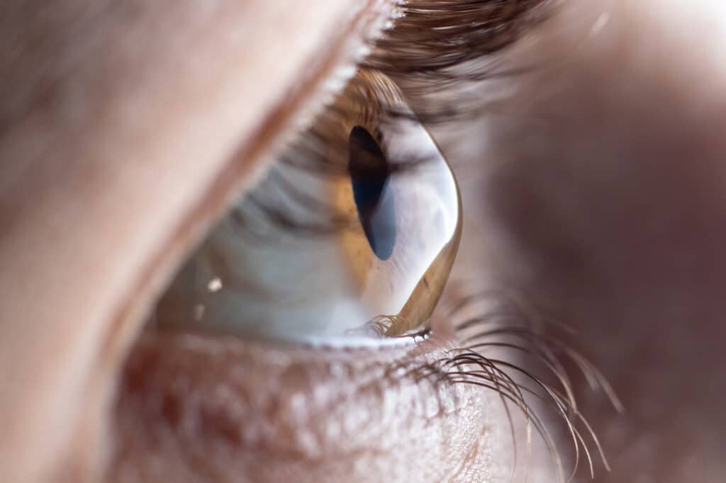
What is Keratoconus?
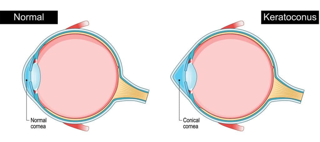
Keratoconus is a condition that causes thinning and bulging of the cornea, the front surface of the eye that allows light to enter. It is a progressive condition that distorts the cornea’s shape. While the condition is present from birth, most people do not experience symptoms until they are between the ages of 10 and 25 years
What are the symptoms of keratoconus?
- Blurred or distorted vision
- Cloudy vision
- Light sensitivity
The distorted cornea shape results in blurred or reduced vision and increased eye sensitivity.
What causes keratoconus?
The exact cause is uncertain, but a genetic link seems likely, as the incidence rate is greater if a family member has been diagnosed. Keratoconus disease affects approximately one person in every 1000. Experiencing seasonal allergies and asthma can be additional risk factors, and the associated rubbing of the eye can make the condition worse.
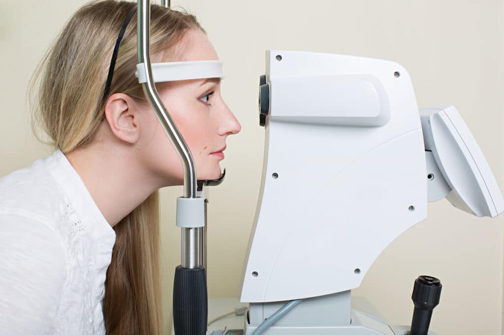
How we diagnose keratoconus
There are a range of vision and corneal measurement assessments we can use to diagnose and monitor your keratoconus symptoms. Refraction testing checks for problems in vision, and uses combinations of different lenses to improve your eyesight. This is the basis for for vision correction corrective glasses. Corneal topography records the shape of the eye’s surface and is used to monitor your condition’s progression – how your eye is changing – over time. Once keratoconus progresses, corneal thickness testing (pachymetry) is another simple and painless way of tracking corneal measurements.
Other Ectasia
Terrien’s Marginal Degeneration
Terrien’s Marginal Degeneration is a progressive and degenerative thinning of the cornea, usually in the superior (upper) margin of the cornea. It is more commonly seen in males over the age of 40. As the condition progresses, the corneal thinning can extend the entire circumference of the cornea. It can be inflammatory in nature, with neovascularisation (blood vessel growth), lipid deposits and corneal scarring commonly seen.
Pellucid Marginal Degeneration
Pellucid Marginal Degeneration is a progressive, degenerative, noninflammatory corneal condition. It is commonly confused with Keratoconus as it also results in thinning of the cornea, however the area of thinning is typically localised to the inferior (lower) and peripheral cornea. Like Keratoconus, patients often present with irregular astigmatism, and vision that can no longer be adequately corrected in glasses or contact lenses.
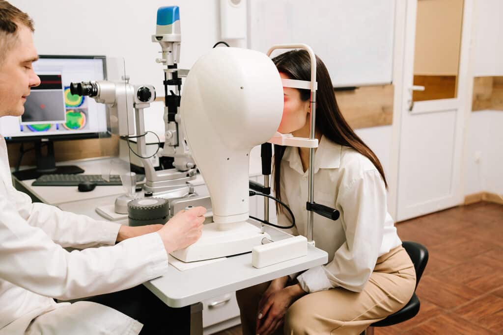
Keratoconus Diagnosis
Keratoconus is diagnosed with a comprehensive eye exam and a discussion of your history of keratoconus disease symptoms. We will carefully analyze the surface and characteristics of your cornea using advanced diagnostic technology. We will also take precise measurements of your cornea to get a baseline idea of its shape, and continue to take annual measurements to detect and track changes.

How we help treat keratoconus
Everyone’s experience of keratoconus is different and the best treatment at one stage will be different to that at another stage. Our highly experienced eye health professionals can provide advice and guidance to suit your needs.
Frequently Asked Questions
Do you have a question or concern about your eye health?
To discuss your condition with an experienced ophthalmologist or optometrist, please contact The Eye Health Centre.